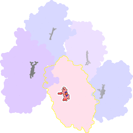
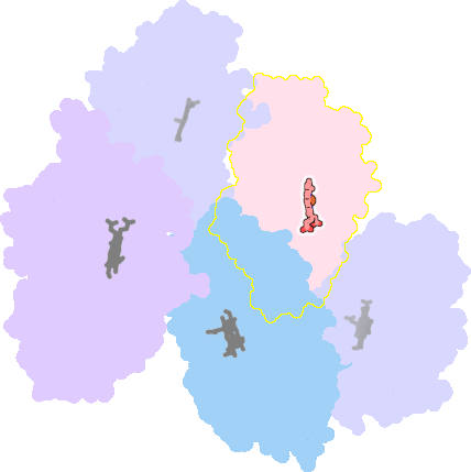
Browser
Protein data
function: oxidoreductaseexperiment: X-RAY DIFFRACTION
resolution: 2.2 Å
origomeric count: 1
axial ligand #1: CYS
chainID: C,resSeq: 347,
Coordination distance[Å]: 2.421
molecule: Vitamin D hydroxylase
Organism: Pseudonocardia autotrophica
List of other hemes in pdb:3a4z
| ID of heme | Distortion | Axial ligands on heme | Function & structure | |
|---|---|---|---|---|
3a4z-A-412 |
sad. -0.28 ruf. -0.47 dom. -0.09 bre. -0.45 |
CYS | chainID: A, resSeq: 347, molecule: Vitamin D hydroxylase |
oxidoreductase oligomeric count: 1 pocket vol.: 409.0 Å3 d(Fe-oop): 0.296 Å |
| Not assigned. | ||||
3a4z-B-412 |
sad. -0.21 ruf. -0.41 dom. -0.08 bre. -0.37 |
CYS | chainID: B, resSeq: 347, molecule: Vitamin D hydroxylase |
oxidoreductase oligomeric count: 1 pocket vol.: 414.0 Å3 d(Fe-oop): 0.297 Å |
| Not assigned. | ||||
3a4z-D-412 |
sad. -0.30 ruf. -0.58 dom. -0.10 bre. -0.35 |
CYS | chainID: D, resSeq: 347, molecule: Vitamin D hydroxylase |
oxidoreductase oligomeric count: 1 pocket vol.: 399.0 Å3 d(Fe-oop): 0.309 Å |
| Not assigned. | ||||
3a4z-E-412 |
sad. -0.31 ruf. -0.34 dom. -0.19 bre. -0.39 |
CYS | chainID: E, resSeq: 347, molecule: Vitamin D hydroxylase |
oxidoreductase oligomeric count: 1 pocket vol.: 398.0 Å3 d(Fe-oop): 0.276 Å |
| Not assigned. | ||||
