Browser
Protein data
function: oxidoreductaseexperiment: X-RAY DIFFRACTION
resolution: 2.5 Å
origomeric count: 13
axial ligand #1: HIS
chainID: A,resSeq: 61,
Coordination distance[Å]: 1.97
molecule: Cytochrome c oxidase subunit 1
EC number: 1.9.3.1
Organism: Bos taurus
axial ligand #2: HIS
chainID: A,resSeq: 378,
Coordination distance[Å]: 1.902
molecule: same as ligand #1
List of other hemes in pdb:2zxw
| ID of heme | Distortion | Axial ligands on heme | Function & structure | |
|---|---|---|---|---|
2zxw-A-516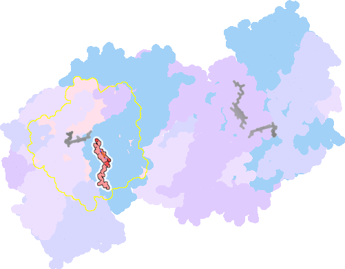 |
sad. -0.39 ruf. -0.13 dom. +0.42 bre. -0.42 |
HIS | chainID: A, resSeq: 376, molecule: Cytochrome c oxidase subunit 1 |
oxidoreductase oligomeric count: 13 pocket vol.: 723.0 Å3 d(Fe-oop): 0.148 Å |
| PER | chainID: A, resSeq: 520, molecule: PEROXIDE ION |
|||
2zxw-N-515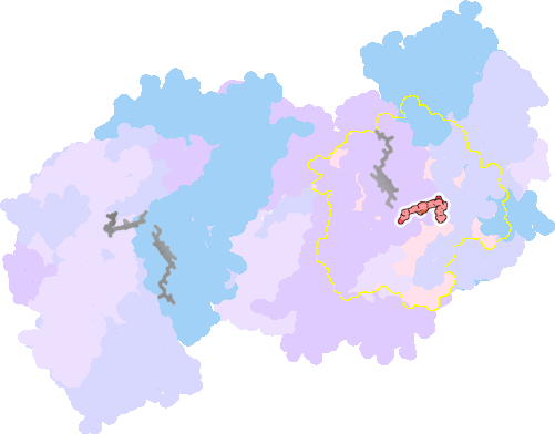 |
sad. -0.57 ruf. -0.04 dom. +0.19 bre. -0.28 |
HIS | chainID: N, resSeq: 61, molecule: Cytochrome c oxidase subunit 1 |
oxidoreductase oligomeric count: 13 pocket vol.: 603.0 Å3 d(Fe-oop): 0.053 Å |
| HIS | chainID: N, resSeq: 378, molecule: Cytochrome c oxidase subunit 1 |
|||
2zxw-N-516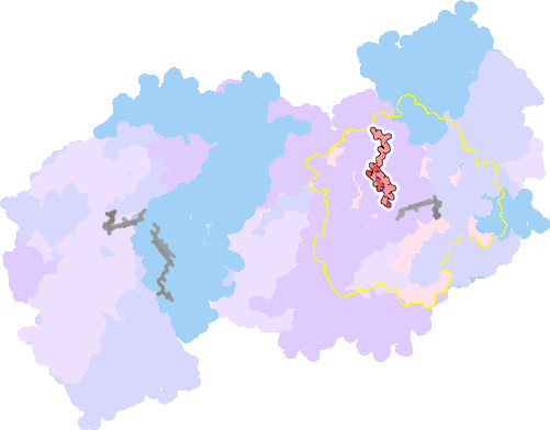 |
sad. -0.23 ruf. -0.06 dom. +0.39 bre. -0.37 |
HIS | chainID: N, resSeq: 376, molecule: Cytochrome c oxidase subunit 1 |
oxidoreductase oligomeric count: 13 pocket vol.: 715.0 Å3 d(Fe-oop): 0.161 Å |
| PER | chainID: N, resSeq: 520, molecule: PEROXIDE ION |
|||