Browser
Protein data
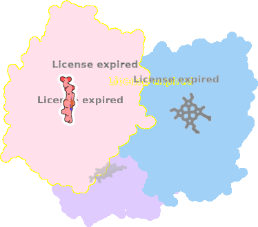
function: oxidoreductaseexperiment: X-RAY DIFFRACTION
resolution: 1.93 Å
origomeric count: 1
axial ligand #1: CYS
chainID: B,resSeq: 377,
Coordination distance[Å]: 2.342
molecule: Steroid C26-monooxygenase
EC number: 1.14.15.29
Organism: Mycobacterium tuberculosis H37Rv
axial ligand #2: DQE
chainID: B,resSeq: 501,
Coordination distance[Å]: 2.591
molecule: ethyl 5-pyridin-4-yl-1~{H}-indole-2-carboxylate
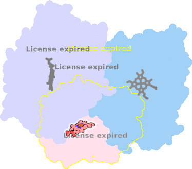
List of other hemes in pdb:7qwn
| ID of heme | Distortion | Axial ligands on heme | Function & structure | |
|---|---|---|---|---|
7qwn-A-502 |
sad. -0.24 ruf. -0.45 dom. -0.15 bre. -0.03 |
CYS | chainID: A, resSeq: 377, molecule: Steroid C26-monooxygenase |
oxidoreductase oligomeric count: 1 pocket vol.: 375.0 Å3 d(Fe-oop): 0.180 Å |
| HOH | chainID: A, resSeq: 654, molecule: water |
|||
7qwn-C-502 |
sad. -0.34 ruf. -0.68 dom. +0.02 bre. -0.12 |
CYS | chainID: C, resSeq: 377, molecule: Steroid C26-monooxygenase |
oxidoreductase oligomeric count: 1 pocket vol.: 389.0 Å3 d(Fe-oop): 0.100 Å |
| DQE | chainID: C, resSeq: 501, molecule: ethyl 5-pyridin-4-yl-1~{H}-indole-2-carboxylate |
|||
