Browser
Protein data
function: lyaseexperiment: X-RAY DIFFRACTION
resolution: 2.3 Å
origomeric count: 1
axial ligand #1: HIS
chainID: B,resSeq: 282,
Coordination distance[Å]: 2.39
molecule: Phenylacetaldoxime dehydratase
EC number: 4.99.1.7
Organism: Bacillus sp. (strain OxB-1)
List of other hemes in pdb:7f2z
| ID of heme | Distortion | Axial ligands on heme | Function & structure | |
|---|---|---|---|---|
7f2z-A-401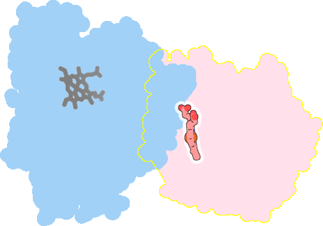 |
sad. -0.44 ruf. -0.26 dom. +0.43 bre. -0.00 |
HIS | chainID: A, resSeq: 282, molecule: Phenylacetaldoxime dehydratase |
lyase oligomeric count: 1 pocket vol.: 500.0 Å3 d(Fe-oop): 0.185 Å |
| Not assigned. | ||||