Browser
Protein data
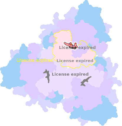
function: electron transporterexperiment: ELECTRON MICROSCOPY
resolution: 2.5 Å
origomeric count: 12
axial ligand #1: HIS
chainID: G,resSeq: 84,
Coordination distance[Å]: 2.774
molecule: Succinate dehydrogenase cytochrome b556 subunit
Organism: Escherichia coli
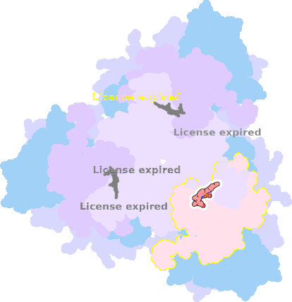
axial ligand #2: HIS
chainID: H,resSeq: 71,
Coordination distance[Å]: 2.818
molecule: Succinate dehydrogenase hydrophobic membrane anchor subunit
Organism: Escherichia coli
List of other hemes in pdb:7jz2
| ID of heme | Distortion | Axial ligands on heme | Function & structure | |
|---|---|---|---|---|
7jz2-D-201 |
sad. -0.81 ruf. +0.32 dom. -0.07 bre. -0.15 |
HIS | chainID: C, resSeq: 84, molecule: Succinate dehydrogenase cytochrome b556 subunit |
electron transporter oligomeric count: 12 pocket vol.: 495.0 Å3 d(Fe-oop): 0.036 Å |
| HIS | chainID: D, resSeq: 71, molecule: Succinate dehydrogenase hydrophobic membrane anchor subunit |
|||
7jz2-K-201 |
sad. -0.81 ruf. +0.32 dom. -0.07 bre. -0.15 |
HIS | chainID: K, resSeq: 84, molecule: Succinate dehydrogenase cytochrome b556 subunit |
electron transporter oligomeric count: 12 pocket vol.: 503.0 Å3 d(Fe-oop): 0.036 Å |
| HIS | chainID: L, resSeq: 71, molecule: Succinate dehydrogenase hydrophobic membrane anchor subunit |
|||
