Browser
Protein data
function: oxygen carrierexperiment: X-RAY DIFFRACTION
resolution: 1.8 Å
origomeric count: 4
axial ligand #1: HIS
chainID: B,resSeq: 92,
Coordination distance[Å]: 2.322
molecule: Hemoglobin subunit beta
Organism: Homo sapiens
axial ligand #2: CMO
chainID: B,resSeq: 148,
Coordination distance[Å]: 1.751
molecule: CARBON MONOXIDE
List of other hemes in pdb:3s65
| ID of heme | Distortion | Axial ligands on heme | Function & structure | |
|---|---|---|---|---|
3s65-A-142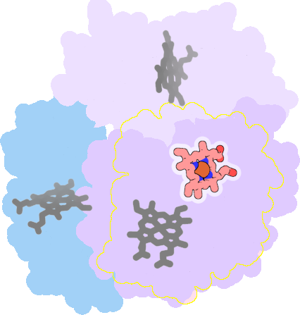 |
sad. +0.31 ruf. -0.35 dom. -0.21 bre. +0.12 |
HIS | chainID: A, resSeq: 87, molecule: Hemoglobin subunit alpha |
oxygen carrier oligomeric count: 4 pocket vol.: 510.0 Å3 d(Fe-oop): 0.124 Å |
| CMO | chainID: A, resSeq: 143, molecule: CARBON MONOXIDE |
|||
3s65-C-142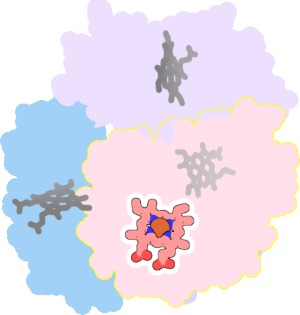 |
sad. +0.14 ruf. -0.30 dom. -0.19 bre. +0.08 |
HIS | chainID: C, resSeq: 87, molecule: Hemoglobin subunit alpha |
oxygen carrier oligomeric count: 4 pocket vol.: 504.0 Å3 d(Fe-oop): 0.131 Å |
| CMO | chainID: C, resSeq: 143, molecule: CARBON MONOXIDE |
|||
3s65-D-147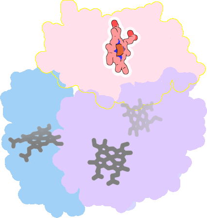 |
sad. +0.14 ruf. -0.47 dom. -0.27 bre. -0.30 |
HIS | chainID: D, resSeq: 92, molecule: Hemoglobin subunit beta |
oxygen carrier oligomeric count: 4 pocket vol.: 558.0 Å3 d(Fe-oop): 0.128 Å |
| CMO | chainID: D, resSeq: 148, molecule: CARBON MONOXIDE |
|||