Browser
Protein data
function: oxidoreductaseexperiment: X-RAY DIFFRACTION
resolution: 1.45 Å
origomeric count: 6
axial ligand #1: HIS
chainID: A,resSeq: 119,
Coordination distance[Å]: 1.974
molecule: Eight-heme nitrite reductase
EC number: 1.7.2.-
Organism: Thioalkalivibrio nitratireducens
axial ligand #2: HIS
chainID: A,resSeq: 300,
Coordination distance[Å]: 1.977
molecule: same as ligand #1
List of other hemes in pdb:3sce
| ID of heme | Distortion | Axial ligands on heme | Function & structure | |
|---|---|---|---|---|
3sce-A-1004 |
sad. +0.53 ruf. -0.49 dom. +0.08 bre. -0.14 |
LYS | chainID: A, resSeq: 188, molecule: Eight-heme nitrite reductase |
oxidoreductase oligomeric count: 6 pocket vol.: 420.0 Å3 d(Fe-oop): 0.030 Å |
| PO4 | chainID: A, resSeq: 1, molecule: PHOSPHATE ION |
|||
3sce-A-1005 |
sad. +1.12 ruf. -0.31 dom. -0.03 bre. +0.00 |
HIS | chainID: A, resSeq: 231, molecule: Eight-heme nitrite reductase |
oxidoreductase oligomeric count: 6 pocket vol.: 534.0 Å3 d(Fe-oop): 0.015 Å |
| HIS | chainID: A, resSeq: 398, molecule: Eight-heme nitrite reductase |
|||
3sce-A-1007 |
sad. +0.77 ruf. -0.64 dom. -0.12 bre. -0.04 |
HIS | chainID: A, resSeq: 383, molecule: Eight-heme nitrite reductase |
oxidoreductase oligomeric count: 6 pocket vol.: 471.0 Å3 d(Fe-oop): 0.030 Å |
| HIS | chainID: A, resSeq: 491, molecule: Eight-heme nitrite reductase |
|||
3sce-A-1008 |
sad. -0.43 ruf. -0.76 dom. +0.17 bre. -0.07 |
HIS | chainID: A, resSeq: 372, molecule: Eight-heme nitrite reductase |
oxidoreductase oligomeric count: 6 pocket vol.: 555.0 Å3 d(Fe-oop): 0.021 Å |
| HIS | chainID: A, resSeq: 415, molecule: Eight-heme nitrite reductase |
|||
3sce-A-1002 |
sad. -0.12 ruf. -0.42 dom. +0.01 bre. -0.12 |
HIS | chainID: A, resSeq: 39, molecule: Eight-heme nitrite reductase |
oxidoreductase oligomeric count: 6 pocket vol.: 384.0 Å3 d(Fe-oop): 0.032 Å |
| HIS | chainID: A, resSeq: 234, molecule: Eight-heme nitrite reductase |
|||
3sce-A-1003 |
sad. +0.35 ruf. -0.55 dom. +0.02 bre. -0.10 |
HIS | chainID: A, resSeq: 30, molecule: Eight-heme nitrite reductase |
oxidoreductase oligomeric count: 6 pocket vol.: 520.0 Å3 d(Fe-oop): 0.035 Å |
| HIS | chainID: A, resSeq: 70, molecule: Eight-heme nitrite reductase |
|||
3sce-A-1001 |
sad. -0.02 ruf. -0.39 dom. -0.05 bre. -0.16 |
HIS | chainID: A, resSeq: 18, molecule: Eight-heme nitrite reductase |
oxidoreductase oligomeric count: 6 pocket vol.: 443.0 Å3 d(Fe-oop): 0.049 Å |
| HIS | chainID: A, resSeq: 44, molecule: Eight-heme nitrite reductase |
|||
3sce-B-1004 |
sad. +0.53 ruf. -0.51 dom. +0.09 bre. -0.15 |
LYS | chainID: B, resSeq: 188, molecule: Eight-heme nitrite reductase |
oxidoreductase oligomeric count: 6 pocket vol.: 397.0 Å3 d(Fe-oop): 0.021 Å |
| HOH | chainID: B, resSeq: 1114, molecule: water |
|||
3sce-B-1005 |
sad. +1.11 ruf. -0.32 dom. -0.04 bre. -0.03 |
HIS | chainID: B, resSeq: 231, molecule: Eight-heme nitrite reductase |
oxidoreductase oligomeric count: 6 pocket vol.: 530.0 Å3 d(Fe-oop): 0.027 Å |
| HIS | chainID: B, resSeq: 398, molecule: Eight-heme nitrite reductase |
|||
3sce-B-1006 |
sad. -0.21 ruf. -0.74 dom. +0.23 bre. -0.07 |
HIS | chainID: B, resSeq: 119, molecule: Eight-heme nitrite reductase |
oxidoreductase oligomeric count: 6 pocket vol.: 412.0 Å3 d(Fe-oop): 0.001 Å |
| HIS | chainID: B, resSeq: 300, molecule: Eight-heme nitrite reductase |
|||
3sce-B-1007 |
sad. +0.77 ruf. -0.63 dom. -0.11 bre. -0.02 |
HIS | chainID: B, resSeq: 383, molecule: Eight-heme nitrite reductase |
oxidoreductase oligomeric count: 6 pocket vol.: 434.0 Å3 d(Fe-oop): 0.028 Å |
| HIS | chainID: B, resSeq: 491, molecule: Eight-heme nitrite reductase |
|||
3sce-B-1008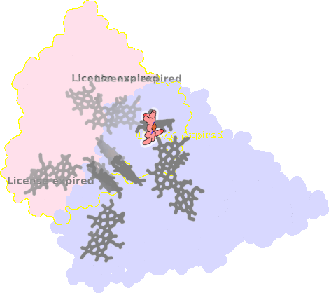 |
sad. -0.41 ruf. -0.77 dom. +0.18 bre. -0.06 |
HIS | chainID: B, resSeq: 372, molecule: Eight-heme nitrite reductase |
oxidoreductase oligomeric count: 6 pocket vol.: 581.0 Å3 d(Fe-oop): 0.014 Å |
| HIS | chainID: B, resSeq: 415, molecule: Eight-heme nitrite reductase |
|||
3sce-B-1002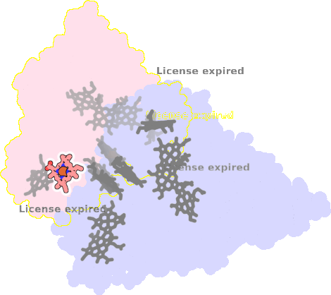 |
sad. -0.13 ruf. -0.44 dom. +0.00 bre. -0.10 |
HIS | chainID: B, resSeq: 39, molecule: Eight-heme nitrite reductase |
oxidoreductase oligomeric count: 6 pocket vol.: 360.0 Å3 d(Fe-oop): 0.022 Å |
| HIS | chainID: B, resSeq: 234, molecule: Eight-heme nitrite reductase |
|||
3sce-B-1003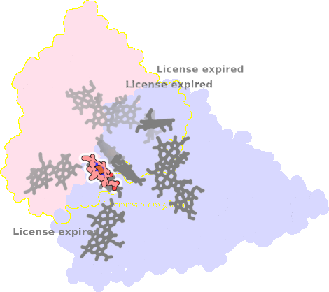 |
sad. +0.34 ruf. -0.50 dom. +0.00 bre. -0.11 |
HIS | chainID: B, resSeq: 30, molecule: Eight-heme nitrite reductase |
oxidoreductase oligomeric count: 6 pocket vol.: 534.0 Å3 d(Fe-oop): 0.022 Å |
| HIS | chainID: B, resSeq: 70, molecule: Eight-heme nitrite reductase |
|||
3sce-B-1001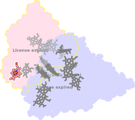 |
sad. -0.22 ruf. -0.36 dom. -0.05 bre. -0.13 |
HIS | chainID: B, resSeq: 18, molecule: Eight-heme nitrite reductase |
oxidoreductase oligomeric count: 6 pocket vol.: 398.0 Å3 d(Fe-oop): 0.033 Å |
| HIS | chainID: B, resSeq: 44, molecule: Eight-heme nitrite reductase |
|||