Browser
Protein data
function: oxidoreductaseexperiment: X-RAY DIFFRACTION
resolution: 1.79 Å
origomeric count: 6
axial ligand #1: HIS
chainID: B,resSeq: 18,
Coordination distance[Å]: 1.902
molecule: Eight-heme nitrite reductase
Organism: Thioalkalivibrio nitratireducens
axial ligand #2: HIS
chainID: B,resSeq: 44,
Coordination distance[Å]: 1.929
molecule: same as ligand #1
List of other hemes in pdb:3s7w
| ID of heme | Distortion | Axial ligands on heme | Function & structure | |
|---|---|---|---|---|
3s7w-A-1004 |
sad. +0.57 ruf. -0.52 dom. +0.09 bre. -0.23 |
LYS | chainID: A, resSeq: 188, molecule: Eight-heme nitrite reductase |
oxidoreductase oligomeric count: 6 pocket vol.: 425.0 Å3 d(Fe-oop): 0.076 Å |
| NO2 | chainID: A, resSeq: 1, molecule: NITRITE ION |
|||
3s7w-A-1005 |
sad. +1.11 ruf. -0.29 dom. +0.00 bre. -0.16 |
HIS | chainID: A, resSeq: 231, molecule: Eight-heme nitrite reductase |
oxidoreductase oligomeric count: 6 pocket vol.: 497.0 Å3 d(Fe-oop): 0.078 Å |
| HIS | chainID: A, resSeq: 398, molecule: Eight-heme nitrite reductase |
|||
3s7w-A-1006 |
sad. -0.20 ruf. -0.72 dom. +0.25 bre. -0.22 |
HIS | chainID: A, resSeq: 119, molecule: Eight-heme nitrite reductase |
oxidoreductase oligomeric count: 6 pocket vol.: 435.0 Å3 d(Fe-oop): 0.010 Å |
| HIS | chainID: A, resSeq: 300, molecule: Eight-heme nitrite reductase |
|||
3s7w-A-1007 |
sad. +0.79 ruf. -0.53 dom. -0.09 bre. -0.15 |
HIS | chainID: A, resSeq: 383, molecule: Eight-heme nitrite reductase |
oxidoreductase oligomeric count: 6 pocket vol.: 440.0 Å3 d(Fe-oop): 0.060 Å |
| HIS | chainID: A, resSeq: 491, molecule: Eight-heme nitrite reductase |
|||
3s7w-A-1008 |
sad. -0.34 ruf. -0.81 dom. +0.23 bre. -0.17 |
HIS | chainID: A, resSeq: 372, molecule: Eight-heme nitrite reductase |
oxidoreductase oligomeric count: 6 pocket vol.: 583.0 Å3 d(Fe-oop): 0.049 Å |
| HIS | chainID: A, resSeq: 415, molecule: Eight-heme nitrite reductase |
|||
3s7w-A-1002 |
sad. -0.15 ruf. -0.41 dom. +0.04 bre. -0.30 |
HIS | chainID: A, resSeq: 39, molecule: Eight-heme nitrite reductase |
oxidoreductase oligomeric count: 6 pocket vol.: 369.0 Å3 d(Fe-oop): 0.064 Å |
| HIS | chainID: A, resSeq: 234, molecule: Eight-heme nitrite reductase |
|||
3s7w-A-1003 |
sad. +0.39 ruf. -0.41 dom. +0.04 bre. -0.24 |
HIS | chainID: A, resSeq: 30, molecule: Eight-heme nitrite reductase |
oxidoreductase oligomeric count: 6 pocket vol.: 535.0 Å3 d(Fe-oop): 0.026 Å |
| HIS | chainID: A, resSeq: 70, molecule: Eight-heme nitrite reductase |
|||
3s7w-A-1001 |
sad. -0.11 ruf. -0.30 dom. -0.08 bre. -0.37 |
HIS | chainID: A, resSeq: 18, molecule: Eight-heme nitrite reductase |
oxidoreductase oligomeric count: 6 pocket vol.: 427.0 Å3 d(Fe-oop): 0.016 Å |
| HIS | chainID: A, resSeq: 44, molecule: Eight-heme nitrite reductase |
|||
3s7w-B-1004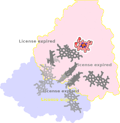 |
sad. +0.61 ruf. -0.44 dom. +0.07 bre. -0.26 |
LYS | chainID: B, resSeq: 188, molecule: Eight-heme nitrite reductase |
oxidoreductase oligomeric count: 6 pocket vol.: 448.0 Å3 d(Fe-oop): 0.067 Å |
| NO2 | chainID: B, resSeq: 1, molecule: NITRITE ION |
|||
3s7w-B-1005 |
sad. +1.10 ruf. -0.29 dom. -0.02 bre. -0.19 |
HIS | chainID: B, resSeq: 231, molecule: Eight-heme nitrite reductase |
oxidoreductase oligomeric count: 6 pocket vol.: 501.0 Å3 d(Fe-oop): 0.046 Å |
| HIS | chainID: B, resSeq: 398, molecule: Eight-heme nitrite reductase |
|||
3s7w-B-1006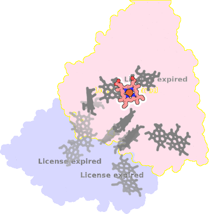 |
sad. -0.16 ruf. -0.73 dom. +0.27 bre. -0.16 |
HIS | chainID: B, resSeq: 119, molecule: Eight-heme nitrite reductase |
oxidoreductase oligomeric count: 6 pocket vol.: 431.0 Å3 d(Fe-oop): 0.039 Å |
| HIS | chainID: B, resSeq: 300, molecule: Eight-heme nitrite reductase |
|||
3s7w-B-1008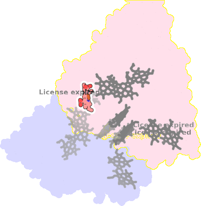 |
sad. -0.32 ruf. -0.77 dom. +0.23 bre. -0.16 |
HIS | chainID: B, resSeq: 372, molecule: Eight-heme nitrite reductase |
oxidoreductase oligomeric count: 6 pocket vol.: 576.0 Å3 d(Fe-oop): 0.084 Å |
| HIS | chainID: B, resSeq: 415, molecule: Eight-heme nitrite reductase |
|||
3s7w-B-1002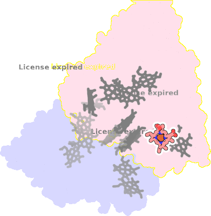 |
sad. -0.12 ruf. -0.40 dom. +0.02 bre. -0.27 |
HIS | chainID: B, resSeq: 39, molecule: Eight-heme nitrite reductase |
oxidoreductase oligomeric count: 6 pocket vol.: 368.0 Å3 d(Fe-oop): 0.045 Å |
| HIS | chainID: B, resSeq: 234, molecule: Eight-heme nitrite reductase |
|||
3s7w-B-1003 |
sad. +0.39 ruf. -0.53 dom. +0.04 bre. -0.22 |
HIS | chainID: B, resSeq: 30, molecule: Eight-heme nitrite reductase |
oxidoreductase oligomeric count: 6 pocket vol.: 518.0 Å3 d(Fe-oop): 0.044 Å |
| HIS | chainID: B, resSeq: 70, molecule: Eight-heme nitrite reductase |
|||