Browser
Protein data
function: electron transporterexperiment: X-RAY DIFFRACTION
resolution: 1.8 Å
origomeric count: 1
axial ligand #1: HIS
chainID: S,resSeq: 38,
Coordination distance[Å]: 2.017
molecule: Cytochrome c554
Organism: Vibrio parahaemolyticus
axial ligand #2: MET
chainID: S,resSeq: 80,
Coordination distance[Å]: 2.393
molecule: same as ligand #1
List of other hemes in pdb:2zzs
| ID of heme | Distortion | Axial ligands on heme | Function & structure | |
|---|---|---|---|---|
2zzs-A-220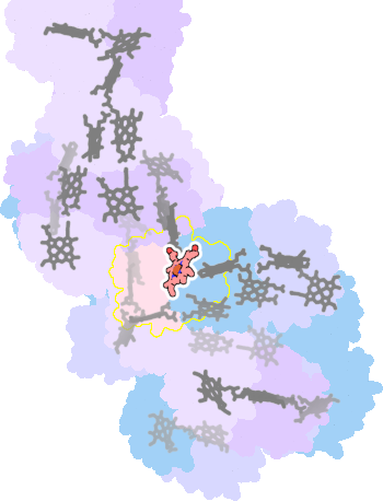 |
sad. -0.02 ruf. -0.61 dom. -0.03 bre. -0.09 |
HIS | chainID: A, resSeq: 38, molecule: Cytochrome c554 |
electron transporter oligomeric count: 1 pocket vol.: 337.0 Å3 d(Fe-oop): 0.025 Å |
| MET | chainID: A, resSeq: 80, molecule: Cytochrome c554 |
|||
2zzs-B-220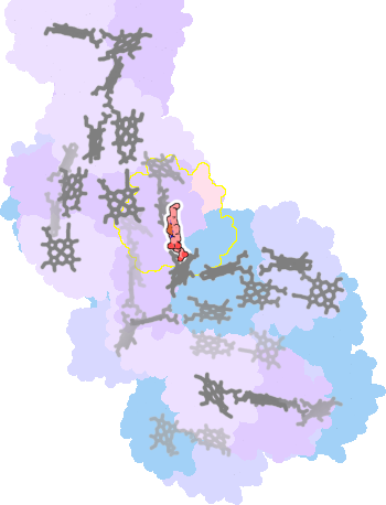 |
sad. +0.11 ruf. -0.43 dom. -0.07 bre. -0.19 |
HIS | chainID: B, resSeq: 38, molecule: Cytochrome c554 |
electron transporter oligomeric count: 1 pocket vol.: 330.0 Å3 d(Fe-oop): 0.011 Å |
| MET | chainID: B, resSeq: 80, molecule: Cytochrome c554 |
|||
2zzs-C-220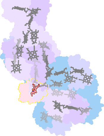 |
sad. +0.03 ruf. -0.35 dom. -0.10 bre. -0.08 |
HIS | chainID: C, resSeq: 38, molecule: Cytochrome c554 |
electron transporter oligomeric count: 1 pocket vol.: 332.0 Å3 d(Fe-oop): 0.027 Å |
| MET | chainID: C, resSeq: 80, molecule: Cytochrome c554 |
|||
2zzs-D-220 |
sad. +0.15 ruf. -0.51 dom. -0.11 bre. -0.19 |
HIS | chainID: D, resSeq: 38, molecule: Cytochrome c554 |
electron transporter oligomeric count: 1 pocket vol.: 360.0 Å3 d(Fe-oop): 0.054 Å |
| MET | chainID: D, resSeq: 80, molecule: Cytochrome c554 |
|||
2zzs-E-220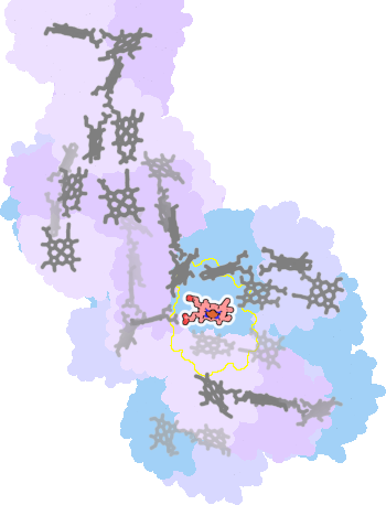 |
sad. +0.13 ruf. -0.44 dom. -0.14 bre. -0.16 |
HIS | chainID: E, resSeq: 38, molecule: Cytochrome c554 |
electron transporter oligomeric count: 1 pocket vol.: 350.0 Å3 d(Fe-oop): 0.033 Å |
| MET | chainID: E, resSeq: 80, molecule: Cytochrome c554 |
|||
2zzs-F-220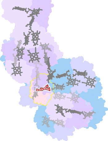 |
sad. +0.10 ruf. -0.47 dom. -0.04 bre. -0.16 |
HIS | chainID: F, resSeq: 38, molecule: Cytochrome c554 |
electron transporter oligomeric count: 1 pocket vol.: 342.0 Å3 d(Fe-oop): 0.065 Å |
| MET | chainID: F, resSeq: 80, molecule: Cytochrome c554 |
|||
2zzs-G-220 |
sad. +0.04 ruf. -0.51 dom. -0.09 bre. -0.24 |
HIS | chainID: G, resSeq: 38, molecule: Cytochrome c554 |
electron transporter oligomeric count: 1 pocket vol.: 362.0 Å3 d(Fe-oop): 0.064 Å |
| MET | chainID: G, resSeq: 80, molecule: Cytochrome c554 |
|||
2zzs-H-220 |
sad. -0.10 ruf. -0.61 dom. -0.04 bre. -0.11 |
HIS | chainID: H, resSeq: 38, molecule: Cytochrome c554 |
electron transporter oligomeric count: 1 pocket vol.: 363.0 Å3 d(Fe-oop): 0.014 Å |
| MET | chainID: H, resSeq: 80, molecule: Cytochrome c554 |
|||
2zzs-I-220 |
sad. +0.02 ruf. -0.35 dom. -0.11 bre. -0.12 |
HIS | chainID: I, resSeq: 38, molecule: Cytochrome c554 |
electron transporter oligomeric count: 1 pocket vol.: 353.0 Å3 d(Fe-oop): 0.018 Å |
| MET | chainID: I, resSeq: 80, molecule: Cytochrome c554 |
|||
2zzs-J-220 |
sad. +0.08 ruf. -0.27 dom. -0.05 bre. -0.20 |
HIS | chainID: J, resSeq: 38, molecule: Cytochrome c554 |
electron transporter oligomeric count: 1 pocket vol.: 364.0 Å3 d(Fe-oop): 0.006 Å |
| MET | chainID: J, resSeq: 80, molecule: Cytochrome c554 |
|||
2zzs-K-220 |
sad. +0.10 ruf. -0.66 dom. +0.01 bre. -0.17 |
HIS | chainID: K, resSeq: 38, molecule: Cytochrome c554 |
electron transporter oligomeric count: 1 pocket vol.: 334.0 Å3 d(Fe-oop): 0.120 Å |
| MET | chainID: K, resSeq: 80, molecule: Cytochrome c554 |
|||
2zzs-L-220 |
sad. +0.07 ruf. -0.32 dom. -0.11 bre. -0.14 |
HIS | chainID: L, resSeq: 38, molecule: Cytochrome c554 |
electron transporter oligomeric count: 1 pocket vol.: 341.0 Å3 d(Fe-oop): 0.061 Å |
| MET | chainID: L, resSeq: 80, molecule: Cytochrome c554 |
|||
2zzs-M-220 |
sad. -0.01 ruf. -0.55 dom. -0.02 bre. -0.14 |
HIS | chainID: M, resSeq: 38, molecule: Cytochrome c554 |
electron transporter oligomeric count: 1 pocket vol.: 369.0 Å3 d(Fe-oop): 0.074 Å |
| MET | chainID: M, resSeq: 80, molecule: Cytochrome c554 |
|||
2zzs-N-220 |
sad. -0.18 ruf. -0.37 dom. -0.04 bre. -0.29 |
HIS | chainID: N, resSeq: 38, molecule: Cytochrome c554 |
electron transporter oligomeric count: 1 pocket vol.: 355.0 Å3 d(Fe-oop): 0.267 Å |
| MET | chainID: N, resSeq: 80, molecule: Cytochrome c554 |
|||
2zzs-O-220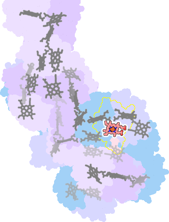 |
sad. +0.01 ruf. -0.61 dom. -0.05 bre. -0.09 |
HIS | chainID: O, resSeq: 38, molecule: Cytochrome c554 |
electron transporter oligomeric count: 1 pocket vol.: 353.0 Å3 d(Fe-oop): 0.067 Å |
| MET | chainID: O, resSeq: 80, molecule: Cytochrome c554 |
|||
2zzs-P-220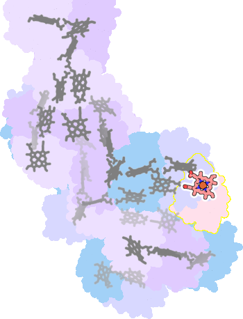 |
sad. -0.01 ruf. -0.33 dom. -0.07 bre. -0.23 |
HIS | chainID: P, resSeq: 38, molecule: Cytochrome c554 |
electron transporter oligomeric count: 1 pocket vol.: 347.0 Å3 d(Fe-oop): 0.097 Å |
| MET | chainID: P, resSeq: 80, molecule: Cytochrome c554 |
|||
2zzs-Q-220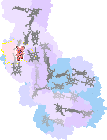 |
sad. -0.12 ruf. -0.50 dom. -0.06 bre. -0.08 |
HIS | chainID: Q, resSeq: 38, molecule: Cytochrome c554 |
electron transporter oligomeric count: 1 pocket vol.: 360.0 Å3 d(Fe-oop): 0.101 Å |
| MET | chainID: Q, resSeq: 80, molecule: Cytochrome c554 |
|||
2zzs-R-220 |
sad. +0.05 ruf. -0.46 dom. -0.16 bre. -0.13 |
HIS | chainID: R, resSeq: 38, molecule: Cytochrome c554 |
electron transporter oligomeric count: 1 pocket vol.: 323.0 Å3 d(Fe-oop): 0.107 Å |
| MET | chainID: R, resSeq: 80, molecule: Cytochrome c554 |
|||
2zzs-T-220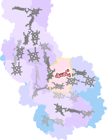 |
sad. -0.04 ruf. -0.33 dom. -0.10 bre. -0.16 |
HIS | chainID: T, resSeq: 38, molecule: Cytochrome c554 |
electron transporter oligomeric count: 1 pocket vol.: 334.0 Å3 d(Fe-oop): 0.009 Å |
| MET | chainID: T, resSeq: 80, molecule: Cytochrome c554 |
|||
2zzs-U-220 |
sad. +0.09 ruf. -0.26 dom. -0.17 bre. -0.18 |
HIS | chainID: U, resSeq: 38, molecule: Cytochrome c554 |
electron transporter oligomeric count: 1 pocket vol.: 347.0 Å3 d(Fe-oop): 0.017 Å |
| MET | chainID: U, resSeq: 80, molecule: Cytochrome c554 |
|||
2zzs-V-220 |
sad. +0.07 ruf. -0.43 dom. -0.16 bre. -0.13 |
HIS | chainID: V, resSeq: 38, molecule: Cytochrome c554 |
electron transporter oligomeric count: 1 pocket vol.: 343.0 Å3 d(Fe-oop): 0.040 Å |
| MET | chainID: V, resSeq: 80, molecule: Cytochrome c554 |
|||
2zzs-W-220 |
sad. -0.06 ruf. -0.54 dom. -0.11 bre. -0.31 |
HIS | chainID: W, resSeq: 38, molecule: Cytochrome c554 |
electron transporter oligomeric count: 1 pocket vol.: 371.0 Å3 d(Fe-oop): 0.019 Å |
| MET | chainID: W, resSeq: 80, molecule: Cytochrome c554 |
|||
2zzs-X-220 |
sad. +0.03 ruf. -0.48 dom. -0.05 bre. -0.20 |
HIS | chainID: X, resSeq: 38, molecule: Cytochrome c554 |
electron transporter oligomeric count: 1 pocket vol.: 361.0 Å3 d(Fe-oop): 0.029 Å |
| MET | chainID: X, resSeq: 80, molecule: Cytochrome c554 |
|||
2zzs-Y-220 |
sad. -0.05 ruf. -0.51 dom. -0.10 bre. -0.19 |
HIS | chainID: Y, resSeq: 38, molecule: Cytochrome c554 |
electron transporter oligomeric count: 1 pocket vol.: 358.0 Å3 d(Fe-oop): 0.132 Å |
| MET | chainID: Y, resSeq: 80, molecule: Cytochrome c554 |
|||
2zzs-Z-220 |
sad. +0.08 ruf. -0.42 dom. -0.08 bre. -0.14 |
HIS | chainID: Z, resSeq: 38, molecule: Cytochrome c554 |
electron transporter oligomeric count: 1 pocket vol.: 375.0 Å3 d(Fe-oop): 0.065 Å |
| MET | chainID: Z, resSeq: 80, molecule: Cytochrome c554 |
|||
2zzs-1-220 |
sad. +0.03 ruf. -0.35 dom. -0.04 bre. -0.09 |
HIS | chainID: 1, resSeq: 38, molecule: Cytochrome c554 |
electron transporter oligomeric count: 1 pocket vol.: 358.0 Å3 d(Fe-oop): 0.011 Å |
| MET | chainID: 1, resSeq: 80, molecule: Cytochrome c554 |
|||
2zzs-2-220 |
sad. +0.02 ruf. -0.45 dom. +0.02 bre. -0.37 |
HIS | chainID: 2, resSeq: 38, molecule: Cytochrome c554 |
electron transporter oligomeric count: 1 pocket vol.: 358.0 Å3 d(Fe-oop): 0.002 Å |
| MET | chainID: 2, resSeq: 80, molecule: Cytochrome c554 |
|||
2zzs-3-220 |
sad. +0.10 ruf. -0.49 dom. -0.02 bre. -0.33 |
HIS | chainID: 3, resSeq: 38, molecule: Cytochrome c554 |
electron transporter oligomeric count: 1 pocket vol.: 361.0 Å3 d(Fe-oop): 0.022 Å |
| MET | chainID: 3, resSeq: 80, molecule: Cytochrome c554 |
|||
2zzs-4-220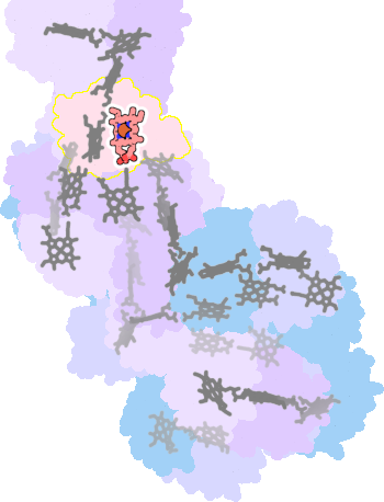 |
sad. -0.04 ruf. -0.50 dom. -0.06 bre. -0.04 |
HIS | chainID: 4, resSeq: 38, molecule: Cytochrome c554 |
electron transporter oligomeric count: 1 pocket vol.: 368.0 Å3 d(Fe-oop): 0.030 Å |
| MET | chainID: 4, resSeq: 80, molecule: Cytochrome c554 |
|||
2zzs-5-220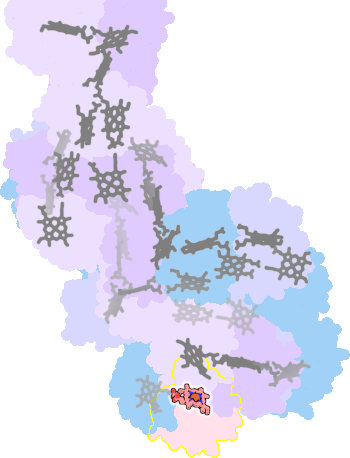 |
sad. +0.20 ruf. -0.93 dom. +0.24 bre. -0.28 |
HIS | chainID: 5, resSeq: 38, molecule: Cytochrome c554 |
electron transporter oligomeric count: 1 pocket vol.: 421.0 Å3 d(Fe-oop): 0.101 Å |
| MET | chainID: 5, resSeq: 80, molecule: Cytochrome c554 |
|||
2zzs-6-220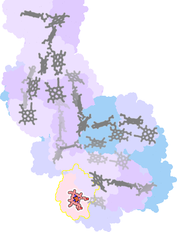 |
sad. +0.25 ruf. -0.96 dom. -0.05 bre. -0.25 |
HIS | chainID: 6, resSeq: 38, molecule: Cytochrome c554 |
electron transporter oligomeric count: 1 pocket vol.: 276.0 Å3 d(Fe-oop): 0.074 Å |
| MET | chainID: 6, resSeq: 80, molecule: Cytochrome c554 |
|||