Browser
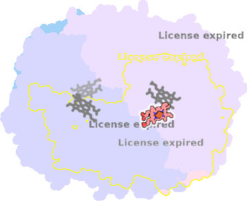
Protein data
function: oxidoreductaseexperiment: X-RAY DIFFRACTION
resolution: 2.28 Å
origomeric count: 4
axial ligand #1: TYR
chainID: B,resSeq: 415,
Coordination distance[Å]: 2.064
molecule: CATALASE HPII
EC number: 1.11.1.6
Organism: Escherichia coli
axial ligand #2: PEO
chainID: B,resSeq: 4002,
Coordination distance[Å]: 2.558
molecule: HYDROGEN PEROXIDE
List of other hemes in pdb:1ggf
| ID of heme | Distortion | Axial ligands on heme | Function & structure | |
|---|---|---|---|---|
1ggf-A-760 |
sad. -0.08 ruf. -0.41 dom. +0.09 bre. -0.03 |
TYR | chainID: A, resSeq: 415, molecule: CATALASE HPII |
oxidoreductase oligomeric count: 4 pocket vol.: 369.0 Å3 d(Fe-oop): 0.353 Å |
| PEO | chainID: A, resSeq: 3001, molecule: HYDROGEN PEROXIDE |
|||
1ggf-C-760 |
sad. -0.03 ruf. -0.46 dom. +0.07 bre. -0.01 |
TYR | chainID: C, resSeq: 415, molecule: CATALASE HPII |
oxidoreductase oligomeric count: 4 pocket vol.: 382.0 Å3 d(Fe-oop): 0.396 Å |
| PEO | chainID: C, resSeq: 5002, molecule: HYDROGEN PEROXIDE |
|||
1ggf-D-760 |
sad. -0.09 ruf. -0.48 dom. -0.05 bre. -0.05 |
TYR | chainID: D, resSeq: 415, molecule: CATALASE HPII |
oxidoreductase oligomeric count: 4 pocket vol.: 354.0 Å3 d(Fe-oop): 0.401 Å |
| Not assigned. | ||||
